MagnetVision™
The MagnetVision™ app is designed to be used as an MR safety teaching tool for those who have attended one of Dr. Emanuel Kanals MRMD/MRSO MR Safety Training Courses. The app represents a graphic embodiment of the Kanal Method, the procedures developed and utilized by Dr. Kanal in his clinical practice when he performs risk assessments on patients with implants/devices/foreign bodies (IDFB) who are requested to undergo MRI examinations.
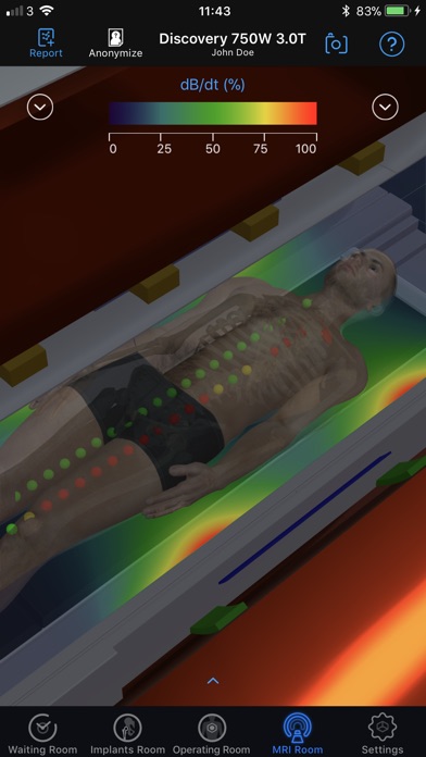
Using spatial field/energy data provided by the manufacturers of the modeled MR scanner hardware, the app functions as a 3D graphic simulator, using colors to visualize the distributions and relative spatial strengths of the invisible energies/magnetic fields present in MRI environments. Photorealistic avatars are customized in real time from user provided gender, height, and weight data to approximate the body habitus of the patient being modeled. 3D implants can be selected/created and labeled with FDA or user defined MR conditions of safety to reflect specific IDFB that may be on/in the patient being modeled. These implants can then be implanted in 3D into the app’s patient avatar in the same position/orientation in which the implant is found in that patient. The avatar is then brought into the MRI Room where an MR scanner/hardware components are selected to be modeled, matching the one to be used in real life in that site. The patient’s avatar is then positioned and centered in the MR scanner just as they would be for the requested MRI examination to be performed. The app then calculates and displays color tracks of the implant as the patient is advanced to the center of the scanner for imaging. If the fields/energies to which the implant will be exposed in the MR imaging process would approach or exceed published/defined maximum safety thresholds for that implant as the patient is advanced into the scanner, the trail and implant would turn yellow and then red, respectively. If the exposure levels to each of these energies/fields is well below published/defined safety thresholds, the implant and trail would be displayed as green.
At the touch of a button the app also produces a detailed, time/date stamped color coded report containing text, graphs, and 3D graphics, documenting the relative field exposure risks of performing the anticipated MR study in the MR scanner being modeled, providing precise feedback and even quantification of exposure levels to the various energies/fields used in the MR imaging process.
Thus, without requiring advanced MR safety expertise on the part of the user, a simple green, yellow, red display for each energy used in the MR environment helps the user instantly identify and even quantify relative risks of proceeding with a requested MR imaging study on a given patient, implant, MR scanner hardware, and MRI examination being modeled. The primary expertise required of the user is to know their patient (gender, height, weight), their implant and its labeling, the MR scanner hardware to be used, and the positioning to be used on that patient’s requested MR study. These are part of the normal expertise of the MR technologist/radiographer, the prime target user of this app. The detailed report generated by the app can then be electronically or hard-copy transmitted to the physician overseeing the MR imaging examination, who can then assess the relative potential risks of such MR exposure while weighing them against the potential benefits of proceeding with the requested MR imaging examination.
The MagnetVision™ app is a ground-breaking superb teaching tool to assess risks associated with MR imaging of IDFB. Users of the app will have taken their first steps towards changing the MR imaging industry by (for most users, perhaps for the very first time) formalizing and standardizing how MR safety risks are assessed, evaluated, and even quantified for patients with implants, devices, and foreign bodies who have been asked to undergo MR imaging examinations.


